Is Your Mole Cancerous?
あなたのほくろは癌でしょうか?
シンガポールで皮膚科の治療を受けられるのはどこでしょうか?
https://singalife.com/category/76399/
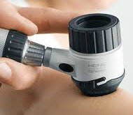
Dermoscopy is a non-invasive diagnostic technique that magnifies the skin in such a way that the colour and structure of the top, middle and lower layers of the upper skin (epidermis, dermoepidermal junction and papillary dermis) becomes visible.
This colour and structure cannot be seen with the naked eye.
Dermoscopy has been shown to significantly increase the clinical diagnosis of skin lesions and with a 30% improvement of diagnosis of melanoma compared to that achieved by clinical examination alone.
Cancerous Mole below as seen under Dermoscopic examination by the doctor:
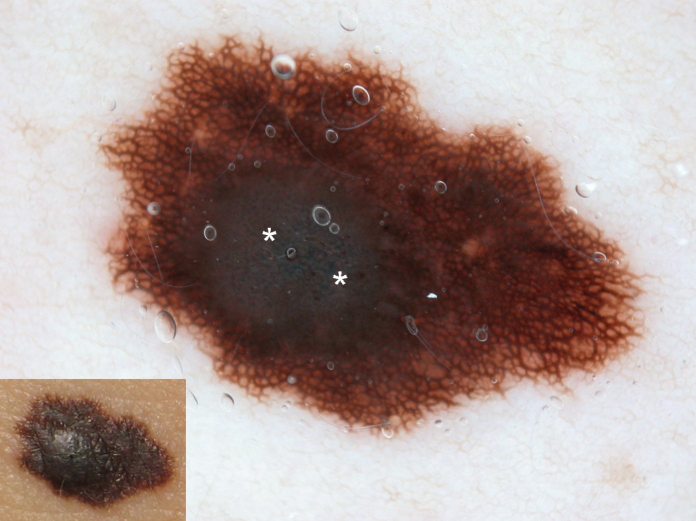
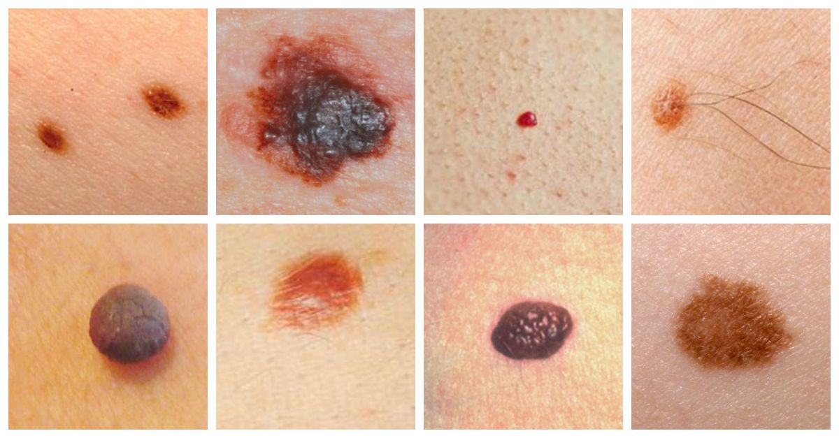
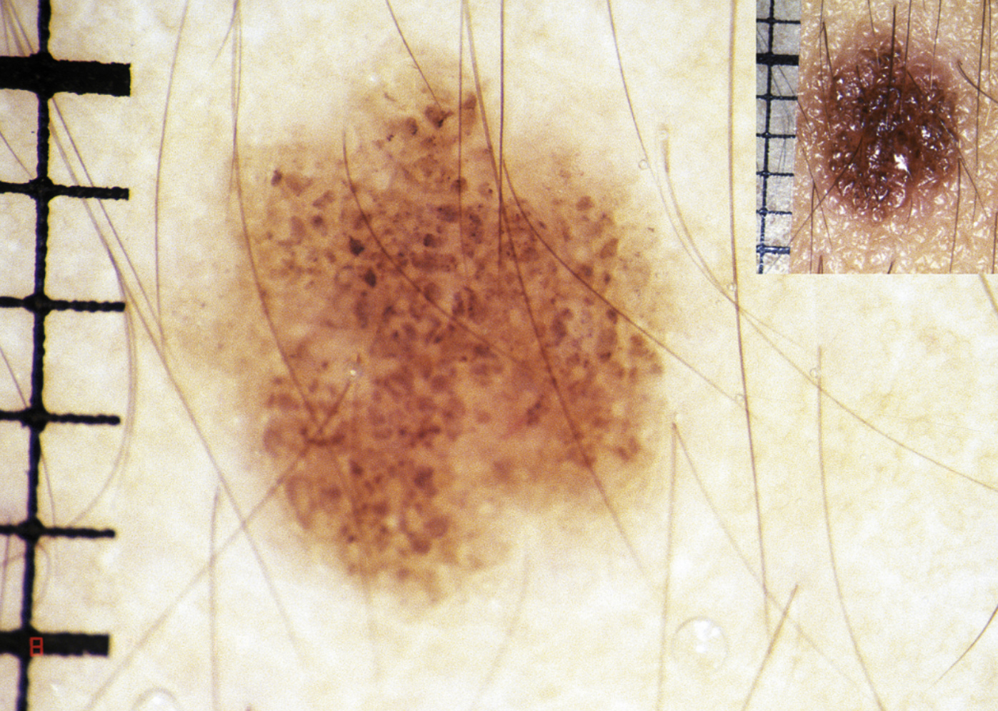
Image below of Benign, non-cancerous moles as seen under a Dermatoscope:
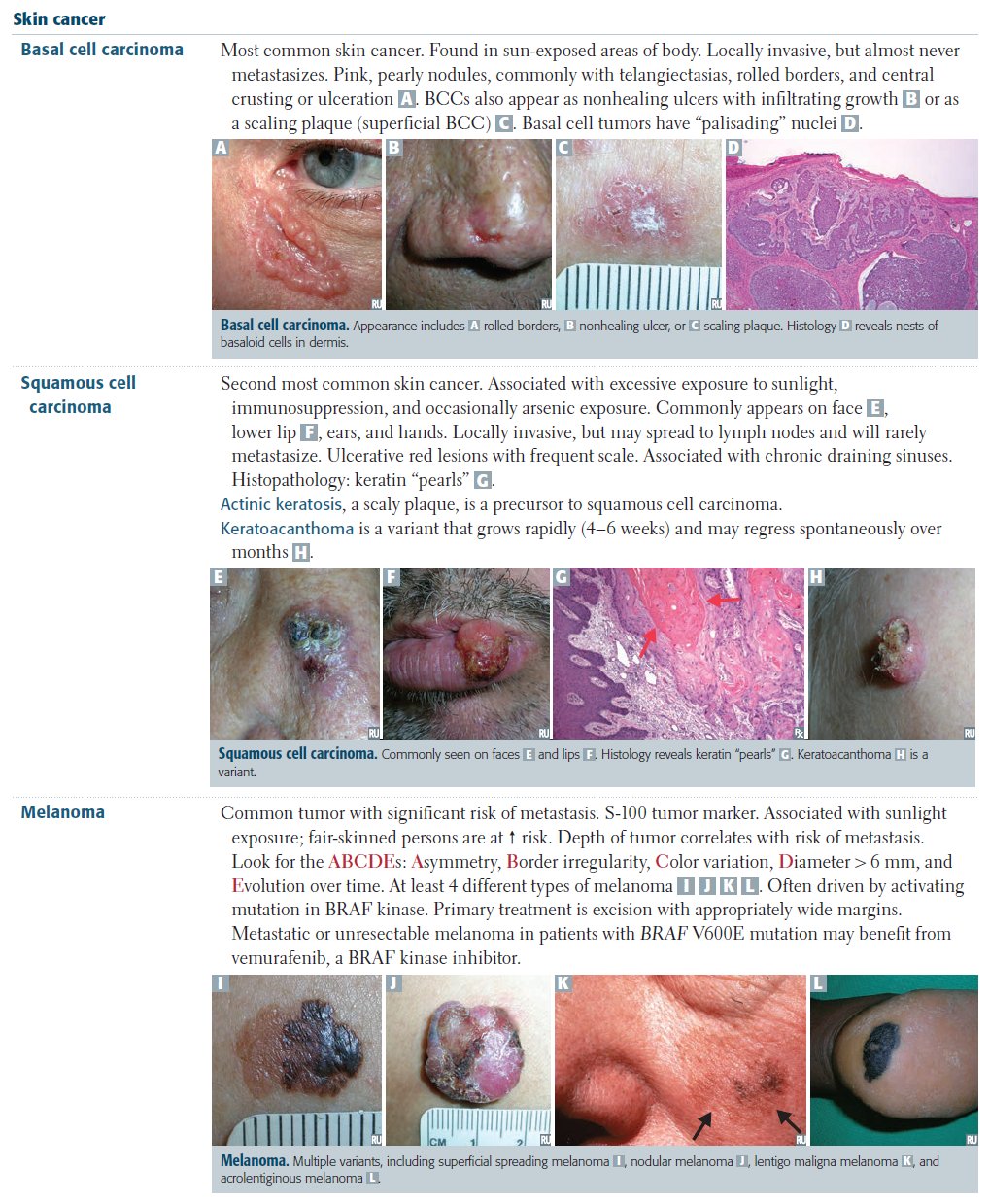
Early detection of a melanoma, basal-cell carcinoma and
squamous-cell carcinoma decreases morbidity and mortality, and therefore result
in better prognosis of malignant skin tumours. The typical application of
dermoscopy is early detection of melanoma.
 |
Dermoscopy is also use to diagnose other types of skin tumours such as basal cell carcinomas, squamous cell carcinomas, cylindromas, dermatofibromas, angiomas, seborrheic keratosis etc. |
It is also used in the diagnosis of scabies and pubic louse. This is achieved by staining the skin with India ink. A dermatoscope can help identify the location of the mite in the burrow, facilitating scraping of the scabetic burrow. The dermatoscope can magnify the very small, difficult to see pubic louse, allowing fast and accurate diagnosis and hence, the treatment.
Suspicious moles can be excised by a doctor for biopsy and histology.
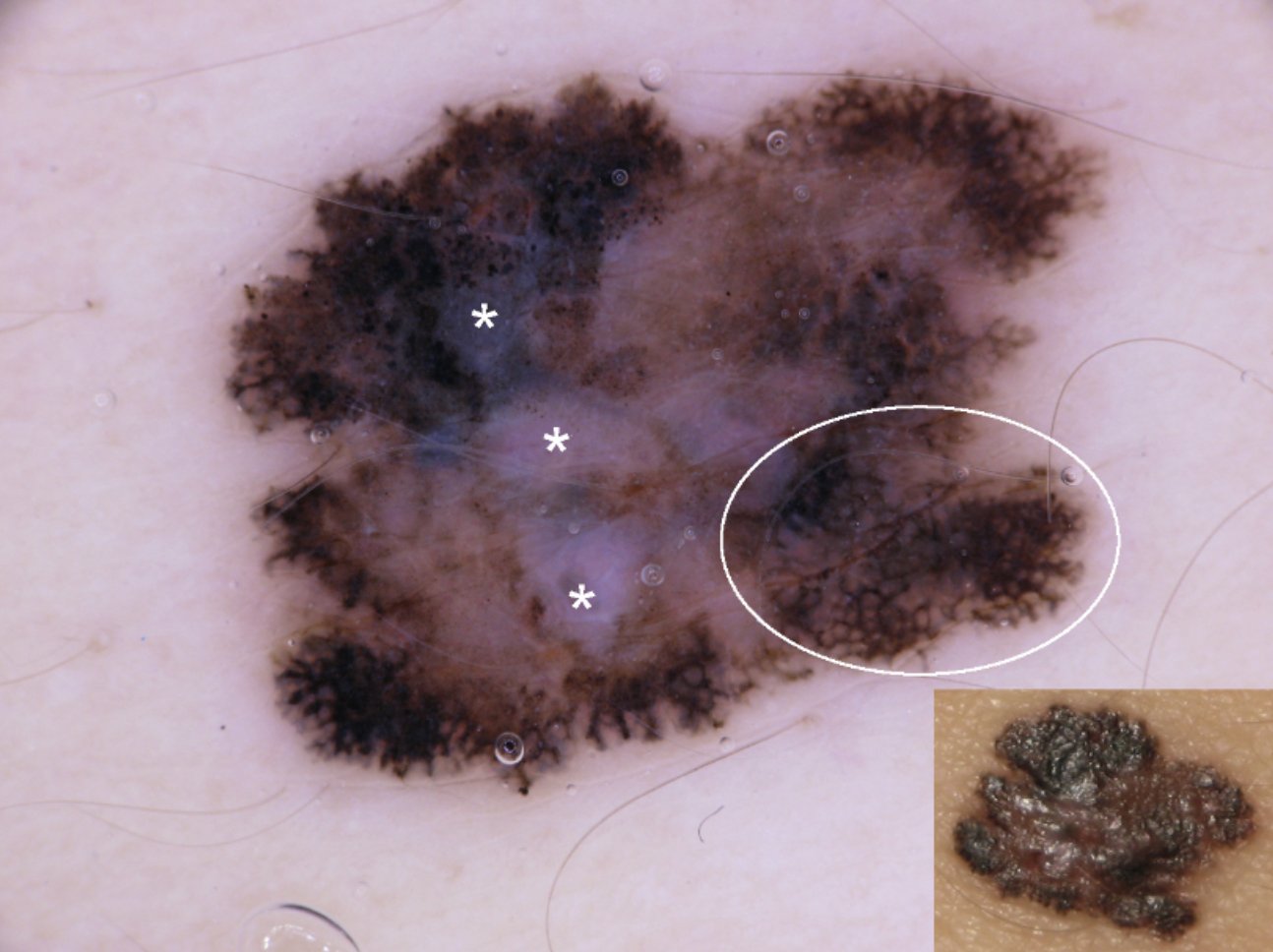
The above picture is of a Cancerous Mole or Melanoma
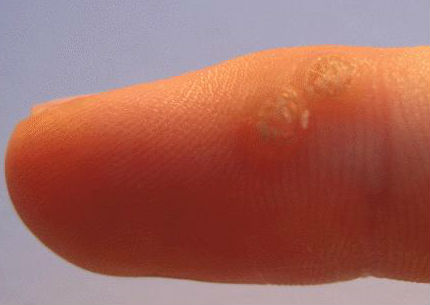
In the treatment of warts, the dermatoscope allows a physician to visualize the structure of a wart and to distinguish it from corn, callouses, trauma, or foreign bodies. Examining warts at later stages of treatment, prevents premature cessation of therapy as the micro-structures of the warts can be follow-up with dermoscopy to ensure if the warts lesion is completely cured.
Dermoscopy of hair and scalp is called trichoscopy. It can help to differentiate the "black dot" of tinea capitis (fungal scalp infection) from alopecia areata.
* The images of cancerous and benign moles are courtesy of the book 'Dermoscopy' and are for educational purposes only. Kindly consult your doctor if you are unsure or suffering from any medical problems.

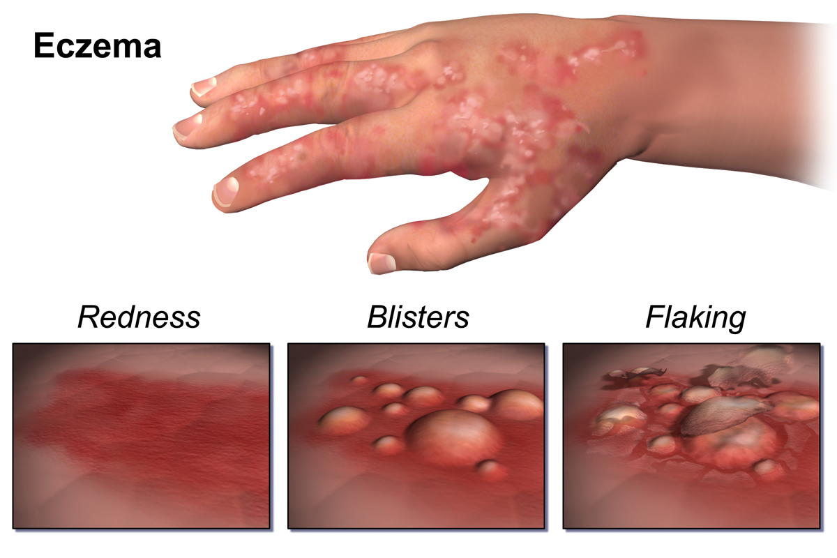
Skin Problems - Dermatology treatment for Allergies, Acne, Warts / Water warts & Eczema

Mina Bissell: Experiments that point to a new understanding of cancer
For decades, researcher Mina Bissell pursued a revolutionary idea -- that a cancer cell doesn't automatically become a tumor, but rather, depends on surrounding cells (its microenvironment) for cues on how to develop. She shares the two key experiments that proved the prevailing wisdom about cancer growth was wrong.
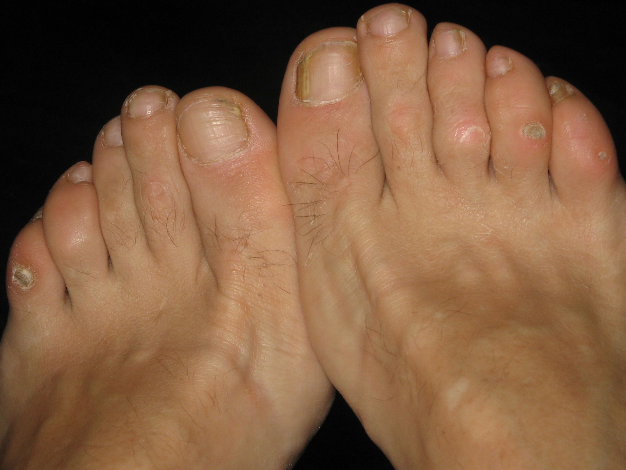
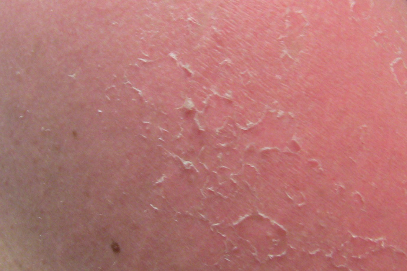
Dermatology Problems - Treatment of Dermatitis, Hives, Warts, Corns & Calluses and Dry Skin

How a Gluten-free Diet can Improve your Chronic Illnesses
Healthy Diets for Optimal Health
|


The information provided in this website is for knowledge purposes only. It does not constitute medical advice.
Should you encounter any medical problem that you are unsure of, always consult your doctor or health care provider for assistance and medical advice.